pregnancyfollowup.com

drsoukainaelaziz@gmail.com

Can Botox Cause Cancer

Can Botox Cause Cancer? A Comprehensive Medical Review of Scientific Evidence

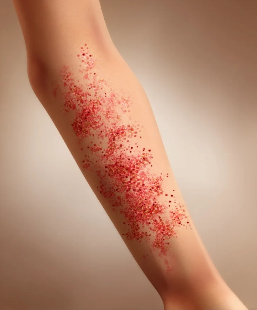
Introduction About Can Botox Cause Cancer The question can Botox cause cancer has become increasingly common among patients considering cosmetic or therapeutic botulinum toxin injections. As Botox continues to be one of the most widely used medical aesthetic treatments worldwide, concerns about its long-term safety—especially potential links to cancer—have grown. These fears are often fueled by misinformation, misunderstanding of the term “toxin,” and anecdotal reports circulating online. Botox has been used in medicine for decades and is approved for both cosmetic and therapeutic indications. Despite its extensive clinical use, some patients remain worried about whether repeated exposure could increase cancer risk. This article provides a detailed, evidence-based medical analysis addressing whether Botox can cause cancer, examining biological mechanisms, clinical studies, epidemiological data, and expert consensus. What Is Botox? Botox is the commercial name for onabotulinumtoxinA, a purified neurotoxin derived from the bacterium Clostridium botulinum. While this bacterium can produce a potent toxin in uncontrolled settings, the form used in medicine is highly purified, standardized, and administered in extremely small, controlled doses. In clinical practice, Botox works by blocking the release of acetylcholine at the neuromuscular junction. This temporarily prevents muscle contraction, leading to muscle relaxation. The effect is localized and reversible, typically lasting three to six months. Botox is widely used for: Given its widespread use, the question can Botox cause cancer deserves a rigorous scientific evaluation. Understanding How Cancer Develops Cancer occurs when normal cells undergo genetic mutations that allow uncontrolled growth, invasion of surrounding tissues, and sometimes metastasis. Known carcinogens typically act through one or more of the following mechanisms: To assess whether Botox can cause cancer, it is essential to determine whether botulinum toxin can induce any of these mechanisms. Biological Plausibility: Can Botox Cause Cancer? From a biological standpoint, Botox does not have characteristics associated with carcinogenic substances. The toxin: Botulinum toxin acts at the synaptic level by cleaving specific proteins involved in neurotransmitter release. This mechanism affects nerve signaling, not cellular replication or genetic stability. Because cancer development requires genetic or epigenetic alterations, there is no plausible biological pathway by which Botox could initiate cancer. This absence of a carcinogenic mechanism strongly argues against the idea that Botox can cause cancer. Clinical Trial Evidence Botox has undergone extensive clinical testing across multiple medical fields. Large randomized controlled trials, observational studies, and post-marketing surveillance data have consistently failed to demonstrate any association between Botox exposure and increased cancer risk. Patients receiving Botox for cosmetic purposes often use it repeatedly over many years. Similarly, patients treated for neurological or urological conditions may receive higher cumulative doses. Despite this long-term exposure, no increase in cancer incidence has been observed compared to the general population. If Botox could cause cancer, such an association would likely have emerged after millions of treatments worldwide. The absence of such findings provides strong reassurance regarding its oncological safety. Long-Term and Epidemiological Data Epidemiological studies are particularly useful for identifying long-term risks such as cancer. Botox has been used in medical practice for over 30 years and in cosmetic medicine for more than two decades. During this time, millions of individuals have been exposed to botulinum toxin. Population-level data do not show higher rates of cancer among Botox users. There is no evidence of increased skin cancer, breast cancer, neurological tumors, or systemic malignancies linked to Botox use. Thus, epidemiological evidence strongly supports the conclusion that Botox does not cause cancer. Laboratory and Preclinical Studies In laboratory studies, botulinum toxin has been examined for potential cytotoxic and mutagenic effects. These studies have consistently shown that Botox does not induce DNA mutations or malignant cellular transformation. Interestingly, some experimental research has explored botulinum toxin for potential therapeutic roles in oncology, such as reducing cancer-related pain or muscle spasm. These investigations further reinforce that Botox does not promote tumor growth. From a preclinical perspective, there is no evidence supporting the idea that Botox can cause cancer. Common Misconceptions About Botox and Cancer The Word “Toxin” One of the main reasons patients ask can Botox cause cancer is fear related to the word “toxin.” In medicine, toxicity depends on dose, route, and context. Many life-saving medications are toxic at high doses but therapeutic at low doses. Botox is administered in microgram quantities and remains localized at the injection site. It is not comparable to environmental toxins or carcinogens. Injection Site Reactions Temporary swelling, redness, or bruising after Botox injections are common and benign. These local inflammatory responses do not indicate cancer and are not associated with malignant transformation. Anecdotal Reports Isolated personal stories claiming cancer after Botox are anecdotal and do not establish causation. Cancer is common in the general population, and coincidental timing does not imply a causal relationship. Botox Side Effects: What Is Actually Known Although Botox does not cause cancer, it is important to acknowledge its known side effects. Common Side Effects Rare but Serious Effects None of these adverse effects involve carcinogenesis or tumor development. Botox Use in Cancer Patients Botox is sometimes used safely in patients with cancer to manage symptoms such as muscle spasticity, chronic pain, or radiation-induced complications. Its use in oncology patients further demonstrates that Botox does not stimulate cancer progression or interfere with cancer treatment. Oncologists do not consider Botox a cancer risk, and it is not contraindicated in cancer survivors when medically appropriate. Regulatory and Expert Consensus Major regulatory agencies worldwide continue to approve Botox for multiple indications. Ongoing pharmacovigilance programs monitor adverse events, including cancer. To date, no regulatory authority has identified cancer as a safety concern associated with Botox. Medical societies in dermatology, neurology, plastic surgery, and aesthetic medicine consistently state that Botox is safe when used appropriately by trained professionals. Final Answer: Can Botox Cause Cancer? Based on decades of scientific research, clinical experience, and population data, the answer to the question can Botox cause cancer is no. There is: Botox remains one of the most studied and safest injectable treatments in modern medicine when administered correctly. Frequently Asked Questions (FAQ) About Can

Can Botox Cause Cancer? A Comprehensive Medical Review of Scientific Evidence Read More »

Can a Prolapse Be a Sign of Cancer

Can a Prolapse Be a Sign of Cancer?

A Medical Perspective on Pelvic Organ Prolapse and Malignancy** Introduction About Can a Prolapse Be a Sign of Cancer Pelvic organ prolapse is a frequent gynecological condition that affects millions of women worldwide, particularly after childbirth and menopause. It occurs when one or more pelvic organs descend from their normal anatomical position due to weakening of the pelvic floor muscles and connective tissues. Because prolapse can cause visible bulging, discomfort, and anxiety, many patients worry about serious underlying causes. A common and understandable question is: can a prolapse be a sign of cancer? The short answer is that a prolapse is not considered a sign of cancer. However, this topic deserves a detailed medical explanation because, in rare situations, prolapse and cancer may coexist, or an underlying malignancy may indirectly contribute to prolapse symptoms. This article provides a comprehensive medical review of the relationship between prolapse and cancer, helping patients and clinicians understand when concern is justified and when reassurance is appropriate. Understanding Pelvic Organ Prolapse Pelvic organ prolapse develops when the support system of the pelvis fails. The pelvic floor is composed of muscles, ligaments, and fascia that hold the bladder, uterus, vagina, rectum, and small bowel in place. When these structures weaken, organs can descend into the vaginal canal or even protrude outside the vaginal opening. Common types of prolapse include: Risk factors include vaginal childbirth, advancing age, menopause, obesity, chronic coughing, constipation, heavy lifting, and prior pelvic surgery. Importantly, none of these factors are directly related to cancer development. Can a Prolapse Be a Sign of Cancer? From a medical standpoint, a prolapse is not a symptom or warning sign of cancer. Pelvic organ prolapse is a mechanical and structural condition rather than a malignant process. Most cases are benign and related to physical stress on pelvic support structures over time. That said, the question can a prolapse be a sign of cancer arises because prolapse can look alarming and because cancers of the pelvis may also cause pelvic symptoms. Understanding the distinction between correlation and causation is essential. Prolapse does not cause cancer, and cancer does not typically cause prolapse. Why Prolapse Is Not Considered a Cancer Symptom Cancer symptoms usually arise from abnormal cellular growth, tissue invasion, bleeding, pain, or systemic effects such as weight loss and fatigue. Prolapse, on the other hand, results from weakened support structures and gravity. The underlying mechanisms are entirely different. In most patients diagnosed with pelvic organ prolapse: Therefore, in routine clinical practice, prolapse alone does not prompt suspicion of cancer. Rare Situations Where Prolapse and Cancer Coexist Although uncommon, medical literature describes rare cases where prolapse and gynecologic cancer occur at the same time. These cases are exceptional and do not change the general rule that prolapse is benign. Cervical Cancer and Uterine Prolapse In elderly women, long-standing uterine prolapse has occasionally been diagnosed alongside cervical cancer. In these cases, cancer was not caused by the prolapse but rather developed independently. The prolapse sometimes delayed diagnosis because symptoms such as discharge or bleeding were mistakenly attributed to mechanical irritation. Vaginal or Endometrial Cancer Rarely, cancers of the vagina or uterus have been discovered during evaluation of advanced prolapse. Again, this does not mean the prolapse was a sign of cancer, but rather that the two conditions coexisted. These rare situations reinforce the importance of thorough gynecological evaluation, especially in postmenopausal women. Indirect Ways Cancer Could Influence Prolapse While answering the question can a prolapse be a sign of cancer, it is important to mention indirect mechanisms: In these cases, prolapse is a consequence of physical changes caused by cancer or its treatment—not a sign of cancer itself. Symptoms That Require Careful Evaluation Although prolapse is usually benign, certain symptoms should never be ignored. These symptoms do not mean cancer is present, but they justify further investigation: When such symptoms are present, clinicians should rule out malignancy through appropriate diagnostic tests. Clinical Assessment and Diagnosis When a patient presents with prolapse, the evaluation typically includes: This approach ensures that serious conditions are not missed while avoiding unnecessary fear. The evaluation is especially important in older women and in those who have not undergone regular gynecologic screening. Does Prolapse Increase Cancer Risk? Current medical evidence does not support the idea that prolapse increases the risk of developing cancer. Chronic exposure of prolapsed tissue can cause irritation or ulceration, but malignant transformation is extremely rare. Therefore, the answer to can a prolapse be a sign of cancer remains no in the vast majority of cases. Prolapse does not predispose women to gynecologic malignancies. Management of Pelvic Organ Prolapse Treatment of prolapse depends on severity, symptoms, and patient preference. Options include: These treatments focus on restoring anatomy and improving quality of life. They do not address cancer because cancer is typically not present. Management When Cancer Is Diagnosed If cancer is detected during prolapse evaluation, management follows standard oncologic protocols. Treatment may include surgery, radiation therapy, chemotherapy, or a combination of approaches. In some cases, prolapse repair and cancer treatment can be planned together, but this requires individualized care by a multidisciplinary team. Patient Reassurance and Education Fear of cancer is common among patients experiencing prolapse, especially when symptoms are visible or uncomfortable. Education is essential. Patients should understand that: Clear communication helps reduce anxiety and supports informed decision-making. Key Points to Remember About Can a Prolapse Be a Sign of Cancer Frequently Asked Questions About Can a Prolapse Be a Sign of Cancer Conclusion About Can a Prolapse Be a Sign of Cancer In conclusion, a prolapse is not a sign of cancer. Pelvic organ prolapse is a common, non-malignant condition caused by weakening of pelvic support structures. While rare cases exist where prolapse and cancer are diagnosed together, these situations are exceptional and should not cause unnecessary alarm. The key message for patients and clinicians is balance: reassurance without complacency. Proper evaluation, attention to warning signs, and routine gynecologic care ensure that serious conditions are identified early while allowing

Can a Prolapse Be a Sign of Cancer? Read More »

Bowel Cancer Stomach Noises

Bowel Cancer Stomach Noises: Medical Explanation, Clinical Significance, and When to Worry

Introduction About Bowel Cancer Stomach Noises Stomach noises are a common bodily phenomenon experienced by people of all ages. These sounds, often described as rumbling, gurgling, or growling, usually reflect normal digestive activity. However, when patients search for information about bowel cancer stomach noises, it often stems from concern that these sounds could indicate a serious underlying condition. While bowel cancer can affect gastrointestinal function, it is essential to understand when stomach noises are harmless and when they may signal a need for medical evaluation. This article provides a comprehensive medical overview of bowel cancer stomach noises, explaining their physiological basis, how bowel cancer may influence digestive sounds, associated symptoms, differential diagnoses, and clinical guidance on when further investigation is necessary. Understanding Stomach Noises: Normal Digestive Physiology Stomach and intestinal noises are medically known as borborygmi. They occur when gas, liquid, and partially digested food move through the gastrointestinal tract. The digestive system relies on rhythmic muscular contractions called peristalsis to push contents forward. These movements naturally produce sound, especially when the intestines contain a mixture of air and fluid. In healthy individuals, stomach noises are most noticeable during fasting, after eating, or following the consumption of certain foods. High-fiber meals, carbonated drinks, and foods that ferment in the gut can all increase gas production and amplify bowel sounds. Importantly, these noises are considered a normal sign of an active digestive system. When discussing bowel cancer stomach noises, it is crucial to recognize that the presence of stomach sounds alone does not indicate disease. In most cases, audible bowel sounds are entirely physiological. How Bowel Cancer Can Affect Digestive Sounds Bowel cancer, also referred to as colorectal cancer, develops in the colon or rectum and may alter normal gastrointestinal function. In certain situations, these changes can influence bowel sounds, leading some patients to notice unusual or persistent noises. Partial Bowel Obstruction As a tumor grows, it may narrow the intestinal lumen. When food, liquid, and gas attempt to pass through a partially obstructed segment, the bowel muscles may contract more forcefully. This increased activity can result in louder or more frequent sounds, sometimes described as high-pitched or exaggerated. In this context, bowel cancer stomach noises may reflect mechanical difficulty in intestinal transit. Altered Intestinal Motility Cancer-related inflammation or disruption of the enteric nervous system can interfere with coordinated peristalsis. This may cause irregular contractions, leading to unpredictable digestive sounds. These altered motility patterns may contribute to sensations of bloating, discomfort, or audible gurgling. Gas Accumulation When bowel contents do not move efficiently, gas can accumulate upstream from a tumor. The movement of trapped gas through narrowed segments may intensify stomach noises. In such cases, bowel cancer stomach noises may be accompanied by abdominal distension or cramping. Despite these mechanisms, it is important to emphasize that bowel cancer does not typically present with stomach noises as an isolated symptom. Common Symptoms of Bowel Cancer To properly interpret bowel cancer stomach noises, they must be considered alongside other, more characteristic symptoms of bowel cancer. These include: When stomach noises occur in combination with these features, especially over several weeks, further medical evaluation becomes essential. Bowel cancer stomach noises gain clinical relevance primarily when associated with these warning signs. When Should Stomach Noises Raise Concern? Most stomach noises are harmless. However, certain characteristics may prompt concern, particularly when evaluating possible bowel cancer stomach noises. Concerning features include: It is the combination of symptoms, rather than the sound itself, that guides clinical suspicion. Patients should be reassured that stomach noises alone are rarely dangerous. Benign Causes Often Confused with Bowel Cancer Many non-cancerous conditions can cause increased bowel sounds, leading patients to worry unnecessarily about bowel cancer stomach noises. Functional Gastrointestinal Disorders Irritable bowel syndrome is a common condition characterized by altered bowel habits, bloating, abdominal discomfort, and audible gut sounds. Unlike bowel cancer, IBS does not cause bleeding, weight loss, or anemia. Food Intolerances and Diet Lactose intolerance, fructose malabsorption, and high-fiber diets can significantly increase gas production. The movement of gas through the intestines commonly produces loud stomach noises without indicating disease. Gastrointestinal Infections Viral or bacterial gastroenteritis often increases bowel motility as the body attempts to expel pathogens. This leads to loud sounds, diarrhea, and cramping that typically resolve within days. Inflammatory Bowel Disease Conditions such as Crohn’s disease or ulcerative colitis can cause chronic inflammation, abdominal pain, diarrhea, and increased bowel sounds. Although these diseases increase cancer risk over time, they are distinct from bowel cancer itself. Understanding these alternatives helps prevent over-attribution of symptoms to bowel cancer stomach noises. Clinical Evaluation and Diagnostic Approach When bowel cancer is suspected, clinicians rely on structured diagnostic pathways rather than symptoms alone. Medical History and Physical Examination The evaluation begins with a detailed discussion of symptoms, duration, family history, and risk factors. Physical examination may reveal abdominal tenderness, masses, or rectal abnormalities. Laboratory Investigations Blood tests are often used to detect anemia, inflammation, or metabolic abnormalities. Stool tests may identify occult blood not visible to the naked eye. Endoscopic Examination Colonoscopy remains the definitive diagnostic tool for bowel cancer. It allows direct visualization of the colon and rectum, as well as biopsy of suspicious lesions. Imaging Studies CT scans or MRI may be used to assess tumor size, spread, or complications such as obstruction. Imaging can also help explain persistent gastrointestinal symptoms, including bowel cancer stomach noises, when structural abnormalities are present. Screening and Prevention Routine screening plays a critical role in reducing bowel cancer mortality. Screening detects precancerous polyps or early-stage cancer before symptoms develop. Individuals over a certain age or with family history should adhere to recommended screening schedules. Lifestyle factors also influence bowel cancer risk. A diet rich in fruits, vegetables, and whole grains, regular physical activity, avoidance of smoking, and moderation of alcohol intake contribute to colorectal health. Patient Education and Reassurance Patients frequently worry that unusual bodily sensations signal serious disease. Education is essential in addressing fears related to bowel cancer stomach noises. Healthcare providers should

Bowel Cancer Stomach Noises: Medical Explanation, Clinical Significance, and When to Worry Read More »

methotrexate ectopic pregnancy​

Methotrexate for Ectopic Pregnancy : A Comprehensive Guide

Introduction About  methotrexate for ectopic pregnancy An ectopic pregnancy occurs when a fertilized egg implants outside the uterine cavity, most commonly within the fallopian tube. Because the tube cannot expand to support the growing pregnancy, ectopic implantation can lead to tube rupture, severe internal bleeding, and life-threatening complications. For this reason, early detection and prompt management are essential. Several treatment options exist depending on the patient’s stability, the size and location of the ectopic pregnancy, and hormone levels. These include expectant management, medical management, and surgery. Among these, methotrexate for ectopic pregnancy has become a widely used non-surgical option that allows preservation of the fallopian tube and future fertility in carefully selected patients. This article provides a detailed overview of how methotrexate works, who qualifies for the treatment, what to expect during therapy, and how it compares to surgical options. What Is Methotrexate? Methotrexate is a medication originally developed as a chemotherapy drug. Over time, it has proven effective in managing several autoimmune and inflammatory conditions, such as rheumatoid arthritis, psoriasis, and certain cancers. Its mechanism of action is based on inhibiting DNA synthesis in rapidly dividing cells. In the context of methotrexate for ectopic pregnancy, the medication targets trophoblastic cells—the cells that rapidly divide to support early pregnancy. By interrupting cell division, methotrexate stops the growth of the ectopic pregnancy and allows the body to gradually reabsorb the pregnancy tissue. Unlike surgery, methotrexate does not physically remove the fallopian tube or damage healthy tissue. This is why it is considered a fertility-preserving option, especially beneficial for women wishing to conceive in the future. Criteria for Methotrexate Use Methotrexate is not suitable for every patient with an ectopic pregnancy. Physicians follow strict criteria to ensure the treatment is both safe and effective. 1. Patient Stability The patient must be hemodynamically stable—meaning normal blood pressure, pulse, and absence of signs of internal bleeding. Methotrexate cannot be used if the fallopian tube has ruptured or if rupture is strongly suspected because surgical intervention becomes urgent. 2. Beta hCG Levels Success rates are highest when initial beta hCG levels are low, typically: Higher hCG levels indicate a more advanced pregnancy and reduced likelihood of methotrexate success. 3. Size and Location of the Ectopic Pregnancy Ultrasound helps evaluate: 4. Contraindications Certain medical conditions make methotrexate unsafe. These include: Patients must also be willing and able to follow through with multiple blood tests and close monitoring. Methotrexate Treatment Protocols Single-Dose Protocol This is the most commonly used protocol. Multi-Dose Protocol This involves alternating methotrexate and leucovorin (a folinic acid rescue agent) across several days. It is typically used when: Because it carries more side effects and requires more monitoring, it is reserved for more complex cases. Monitoring and Follow-Up Even when treatment is successful, hCG levels may take 2 to 6 weeks to return to zero. Follow-up includes: When a Second Dose or Surgery Is Needed A second methotrexate dose may be given if: Surgery becomes necessary when: Effectiveness and Success Rates Methotrexate is generally very effective when used under the right conditions. Success Rate Overall success rates range between 65% and 95%, depending on patient selection. Factors that improve success include: Failure Rates Even with favorable conditions, methotrexate may fail. Around: Failure is more common with advanced ectopic pregnancies or when initial hCG levels are high. Side Effects and Risks Methotrexate is generally safe when administered correctly, but side effects can occur. Common Side Effects Rare but Serious Side Effects Patients should report any concerning symptoms immediately. Warning Signs Requiring Urgent Care These may indicate rupture or heavy internal bleeding: Emergency surgery is required in these cases. Aftercare and Recovery Duration of Recovery It may take several weeks for hCG levels to reach zero. During this period, patients must attend scheduled follow-ups and watch for warning signs. Lifestyle Precautions During treatment with methotrexate for ectopic pregnancy, patients must avoid: Emotional Recovery Ectopic pregnancy can be emotionally distressing. Many women feel grief, anxiety, or fear of recurrence. Supportive counseling or talking with healthcare providers can help. Future Pregnancy Most doctors recommend waiting three months (or two full menstrual cycles) before attempting conception. This ensures: Most women who receive methotrexate go on to have healthy pregnancies later. Comparison with Surgical Treatment Benefits of Methotrexate Limitations Surgical Treatment Options Surgery offers immediate resolution but may reduce fertility if the tube must be removed. Choosing Between the Two The choice depends on: Both methods can be safe and effective when chosen appropriately. Clinical Guidelines and Recommendations Most major medical organizations—including RCOG (UK), NICE, and ACOG (USA)—support methotrexate as a first-line option for selected patients with uncomplicated ectopic pregnancy. Guidelines emphasize: Variations exist between countries depending on available resources, healthcare infrastructure, and professional training. FAQs About  methotrexate for ectopic pregnancy Conclusion About  methotrexate for ectopic pregnancy Methotrexate for ectopic pregnancy is a valuable, non-invasive treatment that helps many women avoid surgery and preserve fertility. Its success depends on early diagnosis, appropriate patient selection, and consistent follow-up. While most women experience a smooth recovery, medical supervision is critical to detect complications such as persistent growth or tubal rupture. Every case is unique. Women diagnosed with ectopic pregnancy should work closely with a specialist to select the safest and most effective management plan for their situation. Early evaluation, clear communication, and timely treatment remain key to ensuring optimal outcomes for both reproductive health and emotional well-being. Written by a Gynecologist based on medical experience and scientific evidence to help you make informed decisions about About methotrexate for ectopic pregnancy

Methotrexate for Ectopic Pregnancy : A Comprehensive Guide Read More »

are hives contagious

Are Hives Contagious? A Medical Perspective

Hives, medically known as urticaria, are a common skin condition that affects people of all ages. They appear as raised, itchy welts that can vary in size, shape, and location on the body. Because hives can look sudden and alarming, patients frequently ask an important question: are hives contagious? Understanding the nature of hives is essential for both patients and healthcare providers, as misconceptions about contagion may lead to unnecessary anxiety, isolation, or fear. From a medical standpoint, the answer to the question are hives contagious is clear: hives themselves are not contagious. They cannot be spread from one person to another through touch, air, or shared objects. However, the conditions that trigger hives may sometimes involve infections that are contagious. This article provides a detailed medical explanation of hives, their causes, mechanisms, and why hives are not considered contagious. Understanding Hives (Urticaria) Hives are a skin reaction characterized by transient wheals, which are raised areas of swelling surrounded by redness. These lesions often itch intensely and may burn or sting. Individual hives typically last less than 24 hours, though new ones may appear as others fade. Hives occur when certain immune cells in the skin, particularly mast cells, release histamine and other inflammatory substances. This causes small blood vessels to leak fluid into surrounding tissues, leading to swelling and redness. Because this reaction originates from within the body, it is not caused by a microorganism spreading from person to person. This is one of the main reasons why the answer to are hives contagious is no. Types of Hives Hives are broadly classified based on their duration and triggers. Acute Urticaria Acute hives last less than six weeks and are the most common form. They often appear suddenly and resolve on their own. Common triggers include: Even in cases where an infection triggers hives, the skin reaction itself does not spread. This distinction is crucial when addressing whether are hives contagious. Chronic Urticaria Chronic hives persist for six weeks or longer and may recur daily or intermittently. In many cases, the exact cause is unknown. Chronic urticaria is often associated with: Chronic hives are never contagious, as they are related to immune system dysregulation rather than infection. Pathophysiology: Why Hives Are Not Contagious To understand why hives are not contagious, it is important to understand how they form. The development of hives is an immunological reaction, not an infectious process. When mast cells are activated, they release histamine, leukotrienes, and other mediators. These substances cause: No bacteria, viruses, or fungi are present in the hives themselves. There is nothing that can be transmitted through contact. Therefore, from a medical and biological perspective, the answer to are hives contagious remains definitively no. Are Hives Ever Related to Contagious Conditions? This is where confusion often arises. While hives themselves are not contagious, some triggers of hives can be contagious. Infection-Related Hives Certain viral or bacterial infections can stimulate the immune system and cause hives as a secondary reaction. Examples include: In these cases: A person exposed to the same infection may develop hives, but this does not mean the hives were transmitted. It means both individuals responded immunologically to the same pathogen. This distinction is essential when clarifying whether are hives contagious. Common Myths About Hives and Contagion Myth 1: You can “catch” hives from someone This is false. Physical contact with a person who has hives will not cause hives in another person. Myth 2: Sharing clothes or bedding spreads hives Hives are not caused by skin parasites or surface organisms. Shared items do not transmit hives. Myth 3: Hives always mean an infection Many cases of hives are allergic or idiopathic and have no infectious cause. Dispelling these myths helps reinforce the medical consensus that are hives contagious is a question with a clear negative answer. Clinical Diagnosis The diagnosis of hives is primarily clinical and based on: Laboratory tests are not usually required for acute hives. In chronic cases, further evaluation may be performed to rule out autoimmune or systemic conditions. Importantly, diagnostic evaluation does not involve infection control measures because hives are not contagious. Treatment and Management Management focuses on symptom relief and trigger avoidance. First-Line Treatment Second-Line Options Advanced Therapies Treatment strategies further emphasize that hives are an internal immune reaction, reinforcing the conclusion that are hives contagious is not a concern in patient management. When to Seek Medical Attention Patients should seek medical care if: These situations do not indicate contagion but may reflect a more serious allergic or systemic reaction. Psychological and Social Considerations Fear of contagion can lead to unnecessary social isolation, especially in children or workplace settings. Educating patients and families that hives are not contagious helps: From a public health and psychological standpoint, answering are hives contagious accurately is essential. Prevention Strategies Although hives cannot be prevented in all cases, certain measures may reduce risk: Preventive strategies do not include isolation or hygiene measures aimed at preventing spread, further supporting the fact that hives are not contagious. Final Answer: Are Hives Contagious? To summarize clearly and medically: Therefore, when patients ask are hives contagious, healthcare providers can confidently reassure them that hives pose no risk of transmission to others. Frequently Asked Questions About Are Hives Contagious Conclusion About Are Hives Contagious Hives are a common, often distressing skin condition, but they are fundamentally non-contagious. The immune-mediated nature of urticaria explains why hives cannot be passed from one person to another. Understanding this distinction is crucial for patient education, proper management, and reducing unnecessary fear. In conclusion, the medical evidence is clear: are hives contagious? No. Accurate information empowers patients, supports appropriate care, and promotes reassurance in both clinical and everyday settings. Written by a Gynecologist based on medical experience and scientific evidence to help you make informed decisions about About Are Hives Contagious

Are Hives Contagious? A Medical Perspective Read More »

Can a Prolapse Be a Sign of Cancer

Is Low Potassium a Sign of Cancer? A Medical Perspective

Introduction About Is Low Potassium a Sign Of Cancer Electrolyte imbalances are common findings in clinical medicine and often raise concern among patients when discovered during routine blood tests. One such imbalance is low potassium, medically referred to as hypokalemia. Because cancer can affect many body systems, patients frequently ask whether abnormal potassium levels may indicate an underlying malignancy. This leads to an important clinical question: is low potassium a sign of cancer? While hypokalemia can occur in patients with cancer, medical evidence shows that low potassium is rarely a direct indicator of malignancy. Instead, it is usually the result of secondary factors such as treatment side effects, nutritional deficiencies, or organ dysfunction. This article provides a detailed medical analysis to clarify whether is low potassium a sign of cancer, explain the physiological role of potassium, review common causes of hypokalemia, and discuss when further investigation is warranted. Understanding Potassium and Its Role in the Body Potassium is an essential electrolyte and mineral that plays a critical role in maintaining normal cellular function. It is primarily an intracellular ion and is vital for: Normal serum potassium levels generally range between 3.6 and 5.0 mmol/L. Levels below this range are classified as hypokalemia. Even mild potassium deficiency can produce symptoms, while severe hypokalemia can lead to life-threatening complications such as cardiac arrhythmias. Because potassium is so essential to vital organs, any persistent abnormality must be evaluated carefully. This often leads patients to wonder is low potassium a sign of cancer, especially when no obvious cause is present. What Is Hypokalemia? Hypokalemia is defined as a serum potassium concentration below the normal reference range. It can be classified based on severity: Symptoms depend on severity and rate of onset. Mild cases may be asymptomatic, while severe cases can cause muscle paralysis, respiratory failure, and dangerous heart rhythm disturbances. Importantly, hypokalemia is a biochemical finding, not a diagnosis in itself. Determining its cause is essential before attributing it to serious conditions such as cancer. Common Causes of Low Potassium Before addressing is low potassium a sign of cancer, it is crucial to understand the far more common non-malignant causes of hypokalemia. These include: 1. Gastrointestinal Losses Vomiting, diarrhea, and chronic laxative use can lead to significant potassium depletion. These are among the most frequent causes of hypokalemia worldwide. 2. Renal Potassium Loss Certain medications, particularly diuretics, increase potassium excretion by the kidneys. Kidney disorders and hormonal imbalances can also promote potassium loss. 3. Inadequate Dietary Intake Low potassium intake, often due to restrictive diets, eating disorders, or poor nutritional status, can contribute to hypokalemia. 4. Hormonal Disorders Conditions such as hyperaldosteronism or Cushing syndrome increase potassium loss through hormonal mechanisms. 5. Intracellular Shifts In some metabolic or endocrine conditions, potassium shifts from the blood into cells, temporarily lowering serum levels without true body depletion. Given these widespread causes, hypokalemia is far more often linked to benign or treatable conditions rather than cancer. Is Low Potassium a Sign of Cancer? The short answer is no, low potassium alone is not a direct or reliable sign of cancer. From a medical standpoint, is low potassium a sign of cancer is best answered by stating that hypokalemia is usually secondary, not primary, in malignant disease. Cancer rarely causes hypokalemia directly. Instead, low potassium may appear in cancer patients due to indirect mechanisms related to the disease or its treatment. How Cancer Can Indirectly Affect Potassium Levels Although is low potassium a sign of cancer is generally answered negatively, there are several indirect pathways through which cancer may contribute to hypokalemia. 1. Cancer Treatment Effects Chemotherapy is a well-recognized cause of electrolyte disturbances. Some chemotherapeutic agents damage renal tubules, leading to increased potassium loss. Others cause nausea, vomiting, or diarrhea, which further deplete potassium stores. Radiation therapy affecting the gastrointestinal tract can also impair nutrient absorption and lead to chronic potassium loss. 2. Reduced Oral Intake Cancer patients may experience loss of appetite, taste changes, or difficulty eating. Reduced potassium intake over time can contribute to hypokalemia, particularly in advanced disease. 3. Paraneoplastic Syndromes In rare cases, tumors produce hormone-like substances that alter electrolyte balance. Certain adrenal or neuroendocrine tumors can cause excessive potassium excretion. In these situations, is low potassium a sign of cancer may be relevant, but such cases are uncommon. 4. Organ Dysfunction Advanced cancers affecting the liver, kidneys, or gastrointestinal system may indirectly disturb potassium regulation due to organ failure. Clinical Evidence and Medical Consensus Medical literature consistently indicates that hypokalemia is not a screening marker for cancer. Studies involving cancer patients show that low potassium is more often associated with: Clinicians do not use potassium levels to diagnose cancer. Instead, potassium is monitored to prevent complications and guide supportive care. Therefore, in the absence of other concerning signs, is low potassium a sign of cancer should not be assumed. Symptoms of Low Potassium Regardless of cause, hypokalemia produces characteristic symptoms, including: These symptoms are nonspecific and occur in many non-cancerous conditions. Their presence alone does not suggest malignancy. When Should Cancer Be Considered? Although uncommon, there are situations where is low potassium a sign of cancer deserves further evaluation: In such cases, additional investigations such as imaging, hormonal assays, or specialist referral may be warranted. Diagnosis and Evaluation of Hypokalemia Evaluation of low potassium includes: Cancer workup is only pursued if clinical findings suggest malignancy beyond electrolyte imbalance. Management of Low Potassium Treatment depends on severity and cause: In cancer patients, proactive electrolyte monitoring is essential during treatment. Correcting potassium imbalance improves quality of life and reduces complications but does not address cancer itself. Key Takeaways Frequently Asked Questions Conclusion About Is Low Potassium a Sign Of Cancer From a medical perspective, is low potassium a sign of cancer is a question that reflects understandable patient concern but is rarely supported by clinical evidence. Hypokalemia is a common electrolyte disturbance with many benign causes. While it may occur in patients with cancer, it is almost always secondary to treatment, nutrition, or

Is Low Potassium a Sign of Cancer? A Medical Perspective Read More »

what level of eosinophils indicate cancer

What Level of Eosinophils Indicate Cancer? A Medical Review for Clinical Understanding

Introduction About What Level of Eosinophils Indicate Cancer Eosinophils are a subtype of white blood cells that play an essential role in immune defense, particularly in allergic reactions, parasitic infections, and inflammatory processes. They are routinely measured as part of a complete blood count with differential. When eosinophil levels are elevated, a condition known as eosinophilia, patients and clinicians alike often ask an important and sometimes anxiety-provoking question: what level of eosinophils indicate cancer? While eosinophilia can be associated with certain malignancies, especially hematologic cancers, it is far more commonly linked to non-malignant conditions. Understanding the clinical significance of eosinophil elevation requires careful interpretation within the broader medical context. This article provides a detailed, medically accurate review of eosinophil levels, their causes, and how they relate to cancer risk, with a focus on evidence-based clinical practice. Understanding Eosinophils and Their Normal Range Eosinophils are granulocytic leukocytes produced in the bone marrow under the influence of cytokines, particularly interleukin-5. Once released into circulation, eosinophils migrate into tissues, especially the lungs, gastrointestinal tract, and skin, where they participate in immune responses. In healthy individuals, eosinophils typically represent 1–6% of circulating white blood cells. The absolute eosinophil count normally ranges between 100 and 500 cells per microliter (µL) of blood. Values above this range are considered abnormal and are categorized as eosinophilia. Eosinophilia is generally classified into three levels: These categories help clinicians assess severity, but they do not directly answer the question of what level of eosinophils indicate cancer. What Level of Eosinophils Indicate Cancer? A crucial medical fact must be stated clearly: there is no specific eosinophil level that definitively indicates cancer. Eosinophil counts alone cannot be used to diagnose malignancy. Instead, eosinophilia may serve as a clinical clue that warrants further evaluation, depending on persistence, severity, and associated symptoms. Most cases of eosinophilia are caused by benign and reversible conditions such as allergies, asthma, parasitic infections, or medication reactions. Cancer-related eosinophilia is relatively rare and usually occurs in specific clinical contexts. Therefore, when patients ask what level of eosinophils indicate cancer, the correct medical answer is that no single numerical threshold confirms cancer, but persistent and unexplained eosinophilia may prompt further investigation. Mechanisms of Eosinophilia in Cancer Eosinophilia associated with cancer can develop through several biological mechanisms: 1. Bone Marrow Involvement In certain hematologic malignancies, such as eosinophilic leukemia or myeloproliferative neoplasms, the bone marrow produces excessive eosinophils due to clonal proliferation. In these cases, eosinophilia is a direct manifestation of the disease. 2. Cytokine-Driven Stimulation Some tumors secrete cytokines, particularly interleukin-5, which stimulate eosinophil production and survival. This indirect mechanism can lead to elevated eosinophil counts even when the cancer does not originate in the bone marrow. 3. Tumor-Associated Immune Response Eosinophils may increase as part of the immune system’s response to malignant cells. This phenomenon reflects immune activation rather than direct tumor production of eosinophils. Types of Cancer Associated With Eosinophilia Hematologic Malignancies The strongest association between eosinophilia and cancer is seen in blood cancers. These include: In these conditions, eosinophil counts are often persistently elevated, sometimes exceeding 1,500 cells/µL, and are accompanied by other abnormal laboratory findings or clinical symptoms. Solid Tumors Eosinophilia is far less common in solid tumors but has been reported in cancers such as: When eosinophilia occurs in solid tumors, it is usually mild to moderate and rarely the only abnormal finding. Importantly, blood eosinophil levels do not reflect tumor burden or stage in most solid cancers. Persistent Eosinophilia and Cancer Risk The duration of eosinophilia is often more clinically relevant than the absolute number. Transient elevations are commonly benign. However, persistent eosinophilia lasting weeks to months, especially when counts exceed 1,500 cells/µL, may raise clinical suspicion. In this context, clinicians reassess the question what level of eosinophils indicate cancer by focusing on: These features do not diagnose cancer but justify deeper evaluation. Symptoms That May Accompany Cancer-Related Eosinophilia Eosinophilia alone is often asymptomatic. However, when related to malignancy, it may coexist with symptoms such as: Organ damage from severe eosinophilia can also occur, affecting the heart, lungs, or nervous system, particularly in chronic eosinophilic disorders. Diagnostic Approach to Elevated Eosinophils Because eosinophilia has many causes, evaluation follows a structured approach: Cancer workup is considered only after common causes are excluded. Thus, even moderate or severe eosinophilia does not automatically answer the question of what level of eosinophils indicate cancer. Prognostic Role of Eosinophils in Cancer Interestingly, eosinophils are not always associated with poor outcomes. In some cancers, higher eosinophil levels or tissue eosinophil infiltration have been linked to: In other malignancies, eosinophilia may reflect aggressive disease biology. Therefore, eosinophils can act as biological markers, but their role varies widely depending on cancer type and clinical setting. Common Non-Cancer Causes of High Eosinophils Understanding benign causes is essential when addressing concerns about what level of eosinophils indicate cancer. Common non-malignant causes include: These conditions account for the vast majority of eosinophilia cases encountered in clinical practice. Key Clinical Takeaways Thus, the question what level of eosinophils indicate cancer cannot be answered by a single number but by comprehensive medical assessment. Frequently Asked Questions (FAQ) About What Level of Eosinophils Indicate Cancer Conclusion About What Level of Eosinophils Indicate Cancer Eosinophils are valuable components of the immune system, and their elevation often reflects benign, treatable conditions. Although eosinophilia can be associated with certain cancers, particularly hematologic malignancies, no specific eosinophil level alone indicates cancer. Persistent, unexplained eosinophilia may warrant further investigation, but it should never be interpreted in isolation. For patients and clinicians alike, understanding the limitations and proper interpretation of eosinophil counts helps prevent unnecessary anxiety while ensuring appropriate medical vigilance. Ultimately, the answer to what level of eosinophils indicate cancer lies not in a single laboratory value, but in thoughtful, evidence-based clinical evaluation. Written by a Gynecologist based on medical experience and scientific evidence to help you make informed decisions about About What Level of Eosinophils Indicate Cancer

What Level of Eosinophils Indicate Cancer? A Medical Review for Clinical Understanding Read More »

13 Week Pregnancy Sonography

13 Week Pregnancy Sonography : What to Expect & Why It Matters

Introduction The 13 week pregnancy sonography is one of the most meaningful moments in early prenatal care. Occurring at the transition between the first and second trimester, this ultrasound scan provides crucial insights into fetal development, maternal health, and future pregnancy planning. At 13 weeks, the embryo has now developed into a fetus with recognizable human features, active movements, and significant organ formation. This scan holds clinical and emotional importance: it reassures parents that the pregnancy is progressing normally, helps healthcare providers evaluate growth and anatomy, and establishes important baselines for future prenatal assessments. Whether this is a woman’s first pregnancy or part of her prenatal routine, the 13-week ultrasound offers clarity, connection, and valuable medical information. Why Is the 13 Week Pregnancy Sonography Done? The 13 week pregnancy sonography serves both diagnostic and evaluative purposes. At this stage, the fetus has undergone rapid development, making it possible for clinicians to perform key measurements and screenings. Confirming Gestational Age and Due Date Although pregnancy dating is often established earlier, the 13-week scan allows for precise measurement of the crown–rump length (CRL), one of the most accurate indicators of gestational age. A corrected due date may be assigned if the measurement significantly deviates from previous estimates. Assessing Fetal Development and Early Anatomy At 13 weeks, the fetus is developed enough for clinicians to inspect major structures, including the skull, limbs, spine, abdomen, and thorax. Any early anomalies, although rare, may be identified. Identifying Multiple Pregnancies Twins or higher-order multiples can be confirmed at this visit, and their chorionicity and amnionicity—critical for managing twin pregnancies—can be determined with high accuracy. Evaluating Placental Health and Amniotic Fluid The placenta’s location, thickness, and general appearance are evaluated. Adequate amniotic fluid volume also provides reassurance that fetal kidneys are functioning and that pregnancy is progressing normally. What Can Be Seen in a 13 Week Pregnancy Sonography ? By 13 weeks, the fetus is about 7.4 cm long, roughly the size of a peach. Thanks to advancements in ultrasound technology, the 13 week pregnancy sonography allows for a wide range of detailed observations. Visible Fetal Anatomy Fetal Movements Even though the mother cannot feel them yet, the fetus is active. Movements may include: These help confirm healthy neuromuscular development. Cardiac Activity and Blood Flow The fetal heart beats between 140–170 bpm at this stage. Using Doppler, the sonographer may observe: Evaluation of the Maternal Cervix and Uterus The cervix is observed to rule out shortening, funneling, or abnormalities. Uterine shape and any fibroids are documented for future follow-up. Nuchal Translucency (NT) Screening during 13 Week Pregnancy Sonography One of the most important parts of the 13 week pregnancy sonography is the Nuchal Translucency (NT) measurement. This test plays a central role in early screening for chromosomal abnormalities. What Is NT? NT refers to the layer of fluid-filled space behind the baby’s neck. At 13 weeks, increased fluid may indicate a higher risk for: Ideal Timing NT can only be accurately measured between: Beyond this window, the measurement is not valid. Normal Range A normal NT measurement is typically below 3 mm, although interpretation also depends on gestational age and fetal length. Additional Factors Used in Risk Calculation The NT measurement does not stand alone. A complete risk assessment includes: Next Steps if Results Are Abnormal An increased NT does not confirm a chromosomal anomaly; it simply indicates elevated risk. Further tests may be recommended: These provide more definitive answers about fetal chromosomal status. Can Gender Be Detected at 13 Week Pregnancy Sonography ? Although still early, determining fetal sex is sometimes possible during the 13 week pregnancy sonography. Nub Theory Between 11–14 weeks, the fetal genital tubercle (nub) may show a directional tilt: Accuracy can exceed 98% when the baby is in an optimal position, but incorrect angles or fetal movement may affect interpretation. However, official confirmation is generally made during the 18–20 week anatomy scan, where visualization is clearer. Preparing for the Ultrasound Proper preparation improves image quality and ensures a smooth appointment. Full Bladder A partially full bladder helps lift the uterus out of the pelvis, providing better imaging—especially for women with a tilted uterus or early scans. Hydration Drinking water throughout the morning improves amniotic fluid clarity, enhancing fetal visibility. Clothing Loose, two-piece outfits make abdominal scanning easier. Bring Essential Documents Emotional Preparedness Bringing a partner or support person may be reassuring. Prepare any questions you may want to ask the sonographer or doctor. What to Expect During the Appointment During the 13 week pregnancy sonography, the sonographer will follow standardized protocols to assess fetal and maternal structures. Type of Scan Procedure Duration Most scans last 45–60 minutes, depending on fetal position and the need for additional views. Results Some clinics provide results immediately, while others deliver reports within 1–3 days. Emotional & Medical Significance of the 13 Week Pregnancy Sonography The 13 week pregnancy sonography is more than a clinical examination—it is a milestone. For many parents, seeing the baby’s movements, hearing the heartbeat, and observing tiny details creates deep emotional bonding. Medically, this scan provides the foundation for risk assessment, pregnancy planning, and ongoing prenatal monitoring. Early identification of abnormalities—though rare—allows for timely follow-up, genetic counseling, and appropriate decisions. Conclusion The 13 week pregnancy sonography is a key milestone that marks the shift into the second trimester and provides essential insight into both fetal and maternal health. This ultrasound helps confirm normal growth, evaluate early anatomy, assess chromosomal risk factors through NT screening, and establish the foundation for the rest of the pregnancy. Beyond its medical importance, this scan offers reassurance and emotional connection, allowing parents to see their baby’s movements and development for the first time. With regular prenatal checkups, open communication with healthcare providers, and a good understanding of each pregnancy stage, women can make confident decisions that support a healthy pregnancy and a positive journey toward childbirth. FAQs Written by a Gynecologist based on medical experience and scientific evidence to help you make informed decisions about 13 Week Pregnancy Sonography

13 Week Pregnancy Sonography : What to Expect & Why It Matters Read More »

Vulvar Melanoma

Vulvar Melanoma : Symptoms, Diagnosis, Treatment, and What Women Should Know

Introduction Melanoma is an aggressive form of cancer that originates in pigment-producing cells known as melanocytes. While most people associate melanoma with the skin exposed to sunlight, it can also develop in mucosal areas of the body where sunlight is not a factor. One of the rarest and most challenging forms is vulvar melanoma, a type of cancer that develops on the external female genitalia. Although uncommon, this form of melanoma is particularly dangerous due to its tendency to be diagnosed at a later stage. Many women may overlook early symptoms or confuse them with benign vulvar conditions, which delays medical attention. Understanding this disease—its symptoms, risk factors, diagnostic steps, and treatment options—is essential for early detection and improved outcomes. Because vulvar melanoma does not behave like more common skin melanomas, awareness among both clinicians and women is crucial. What Is Vulvar Melanoma? Vulvar melanoma arises from melanocytes located within the vulva, including the labia majora, labia minora, clitoris, and perineal regions. These melanocytes produce melanin, the pigment responsible for skin color. Unlike melanoma that appears on sun-exposed skin, vulvar melanoma is classified as a mucosal melanoma, which behaves differently, spreads more aggressively, and often presents diagnostic challenges. It is important to distinguish vulvar melanoma from other types of cancers that affect the vulva. The majority of vulvar cancers—about 70%—are squamous cell carcinomas, which originate from the squamous cells covering the vulvar skin. Vulvar melanoma, in contrast, represents a much smaller percentage of cancers in this region but carries a more serious prognosis. Vulvar melanoma also differs from cutaneous melanoma in its underlying causes, progression pattern, and response to treatment. Because the vulva is not frequently examined and changes may not be easily seen, many women do not notice abnormalities until the tumor has grown significantly. Prevalence and Demographics Although melanoma is a common cancer overall, vulvar melanoma is extremely rare. It accounts for less than 10% of all vulvar cancers and only about 1% of all melanomas in women. Despite its rarity, the disease is clinically significant because of its aggressive nature and low survival rates compared to cutaneous melanomas. Vulvar melanoma is most frequently diagnosed in postmenopausal women, with a median age of around 68 years at diagnosis. However, it can occur at any age and should not be dismissed in younger patients. While more common among white women, this cancer can affect women of all ethnic backgrounds. Because there is no routine screening test for vulvar melanoma, awareness and self-examination are particularly important in older women who may already be experiencing vulvar atrophy or other age-related changes that mask symptoms. Causes and Risk Factors The exact cause of vulvar melanoma remains unclear. Unlike cutaneous melanoma, which is strongly associated with ultraviolet radiation from the sun, mucosal melanomas—including vulvar melanoma—do not share this environmental risk factor. Researchers and clinicians have identified several potential risk factors that may contribute to the development of vulvar melanoma: Age and Menopausal Status Risk increases significantly with age, especially after menopause. Hormonal changes may play a role, although the exact mechanisms are not fully understood. Personal or Family History of Melanoma Women with a history of melanoma elsewhere on the body, or with a close family member affected by melanoma, may have a higher risk. Atypical Moles (Dysplastic Nevi) Although less common in mucosal areas, atypical moles may indicate a predisposition to melanoma. Genetic Factors Studies show potential associations with mutations in genes such as KIT, NRAS, and BRAF, although their impact differs from cutaneous melanoma. Research is ongoing to better understand these molecular pathways. Despite these identified factors, many women diagnosed with vulvar melanoma have no known risk factors, making awareness of symptoms even more critical. Symptoms One of the biggest challenges with vulvar melanoma is that early symptoms are often subtle. Many women attribute discomfort or discoloration to benign conditions such as yeast infections, dermatitis, or normal aging changes. This delay can allow the tumor to progress. Common symptoms of vulvar melanoma include: Itching (Pruritus) Persistent or unexplained itching is one of the most frequently reported early signs. Bleeding Any bleeding not related to menstruation or trauma should be evaluated. Discoloration Lesions may appear black, brown, blue, red, pink, or even white. Because vulvar melanoma can be amelanotic (non-pigmented), unusual pink or red nodules should not be ignored. Pain or Tenderness Pain may appear in later stages but can sometimes occur early. Lumps or Nodules A new growth, swelling, or raised area may indicate tumor development. Ulceration or Open Sores Non-healing ulcers are a concerning sign and require immediate evaluation. Monitoring Lesions Using the ABCDE Rule Although developed for skin melanoma, the ABCDE rule can help identify suspicious vulvar lesions: Women should seek medical evaluation for any persistent change lasting more than two weeks. Diagnosis Vulvar melanoma cannot be detected through a Pap smear, which only screens for cervical abnormalities. Therefore, diagnosis relies on clinical assessment and targeted evaluation of suspicious lesions. Physical Examination A gynecologist or dermatologist examines the vulva carefully for pigmentation changes, ulcers, nodules, or irregular lesions. Biopsy (Gold Standard) A biopsy is essential for confirming a diagnosis of vulvar melanoma. This may involve incisional, excisional, or punch biopsy depending on the lesion’s size and location. Pathology and Breslow Thickness The pathologist evaluates: The Breslow thickness measurement is one of the most important prognostic factors. Thicker tumors have a higher risk of metastasis. Imaging for Staging To determine the extent of the disease, clinicians may use: These tests help identify lymph node involvement or distant metastasis, which guide therapeutic decisions. Stages and Prognosis Staging vulvar melanoma follows general melanoma staging principles, focusing on: Prognostic Value of Breslow Thickness Thin tumors (<1 mm) have a much better prognosis than those with deeper invasion. Survival Rates Although survival depends on many individual factors, data show: Recurrence is unfortunately common, even after aggressive treatment, emphasizing the need for lifelong monitoring. Treatment Options Treatment is tailored based on the stage of the cancer, overall health, and tumor characteristics. Vulvar melanoma requires

Vulvar Melanoma : Symptoms, Diagnosis, Treatment, and What Women Should Know Read More »

7 foods for shrinking fibroids

7 Foods for Shrinking Fibroids: An Evidence-Based Medical Perspective

Uterine fibroids, also known as leiomyomas, are benign tumors originating from the smooth muscle cells of the uterus. They are one of the most common gynecological conditions affecting women of reproductive age. Although fibroids are non-cancerous, they can cause significant symptoms, including heavy menstrual bleeding, pelvic pain, pressure symptoms, anemia, infertility, and recurrent pregnancy loss. Fibroid growth is influenced by hormonal, genetic, and environmental factors, particularly estrogen and progesterone levels. In recent years, growing attention has been given to lifestyle and dietary interventions as complementary approaches to fibroid management. While medical and surgical treatments remain essential in many cases, nutrition may play a supportive role in slowing fibroid growth, reducing symptoms, and improving overall hormonal balance. This article explores 7 foods for shrinking fibroids, focusing on their biological mechanisms, nutritional value, and relevance in fibroid-friendly dietary patterns. Understanding the Relationship Between Diet and Fibroids Fibroids are hormonally responsive tumors. Estrogen promotes fibroid cell proliferation, while progesterone supports fibroid growth and extracellular matrix production. Diet influences estrogen metabolism, inflammation, oxidative stress, and insulin resistance, all of which are implicated in fibroid development. A diet rich in fiber, antioxidants, and anti-inflammatory compounds may support estrogen detoxification through the liver and gastrointestinal tract. Conversely, diets high in red meat, processed foods, and refined carbohydrates have been associated with increased fibroid risk. Understanding this relationship provides the foundation for selecting the 7 foods for shrinking fibroids discussed below. 1. Leafy Green Vegetables Leafy green vegetables such as spinach, kale, Swiss chard, and collard greens are fundamental components of a fibroid-supportive diet. These vegetables are rich in dietary fiber, folate, magnesium, and antioxidants. Fiber plays a critical role in estrogen metabolism by binding estrogen in the intestines and promoting its excretion, thereby reducing circulating estrogen levels. Since fibroid growth is estrogen-dependent, this mechanism is particularly relevant. Leafy greens also provide antioxidants that help reduce oxidative stress and chronic inflammation, both of which contribute to fibroid growth and symptom severity. Regular consumption of leafy greens supports liver function and overall metabolic health. Including leafy greens daily is one of the simplest and most effective strategies when focusing on 7 foods for shrinking fibroids. 2. Berries Berries such as blueberries, strawberries, raspberries, and blackberries are nutrient-dense fruits with a high concentration of antioxidants, including flavonoids and anthocyanins. Oxidative stress has been implicated in abnormal cell proliferation, including fibroid tissue growth. The antioxidants found in berries help neutralize free radicals and reduce inflammatory pathways that may stimulate fibroid progression. In addition to their antioxidant properties, berries are high in fiber and low in glycemic load, making them beneficial for insulin regulation. Insulin resistance has been linked to increased estrogen production in adipose tissue, which may indirectly influence fibroid growth. As part of the 7 foods for shrinking fibroids, berries offer a natural, low-sugar option to support hormonal balance and cellular health. 3. Whole Grains Whole grains such as brown rice, quinoa, oats, barley, and whole wheat are superior alternatives to refined grains when managing fibroids. These foods are rich in complex carbohydrates, fiber, B vitamins, and essential minerals. Fiber from whole grains supports estrogen elimination and improves gut health, both critical for hormonal regulation. Whole grains also help maintain stable blood glucose levels, reducing insulin spikes that may contribute to hormonal imbalances. Replacing refined carbohydrates with whole grains supports weight management, which is important because excess body fat increases estrogen production. A whole-grain-based diet is therefore a logical component of the 7 foods for shrinking fibroids approach. 4. Cruciferous Vegetables Cruciferous vegetables include broccoli, cauliflower, Brussels sprouts, cabbage, and bok choy. These vegetables are particularly important for estrogen metabolism. They contain glucosinolates, which are converted in the body into biologically active compounds such as indole-3-carbinol. These compounds promote the formation of less potent estrogen metabolites and enhance liver detoxification pathways. By supporting healthy estrogen metabolism, cruciferous vegetables may help reduce hormonal stimulation of fibroid tissue. They also provide fiber, vitamins, and anti-inflammatory nutrients that support overall reproductive health. Including cruciferous vegetables regularly strengthens the scientific rationale behind 7 foods for shrinking fibroids. 5. Flaxseeds Flaxseeds are one of the most studied foods in relation to hormone-dependent conditions. They are rich in lignans, a type of phytoestrogen that can modulate estrogen activity in the body. Lignans bind to estrogen receptors and may exert a mild anti-estrogenic effect in high-estrogen environments. This property makes flaxseeds particularly relevant for fibroid management. Flaxseeds also promote favorable estrogen metabolism and increase the production of protective estrogen metabolites. Additionally, flaxseeds are an excellent source of fiber and omega-3 fatty acids, both of which contribute to reduced inflammation and improved hormonal balance. Because of their unique hormonal effects, flaxseeds are a key element of the 7 foods for shrinking fibroids. 6. Turmeric Turmeric is a spice widely recognized for its anti-inflammatory and antioxidant properties. Its active compound, curcumin, has been studied for its effects on cell proliferation and inflammation. Inflammation plays a role in fibroid growth and symptom severity. Curcumin has been shown in laboratory studies to inhibit fibroid cell proliferation and promote programmed cell death. Although large-scale human trials are limited, the biological mechanisms support turmeric’s inclusion in fibroid-supportive diets. Turmeric may also support liver detoxification and reduce oxidative stress, further contributing to hormonal balance. When discussing 7 foods for shrinking fibroids, turmeric stands out as a functional food with therapeutic potential. 7. Healthy Fats Healthy fats from sources such as olive oil, avocados, nuts, and seeds are essential for hormone production and inflammation control. These fats contain monounsaturated and omega-3 fatty acids that help regulate inflammatory pathways and support cell membrane integrity. Chronic inflammation can worsen fibroid symptoms, including pain and heavy bleeding. Healthy fats also play a role in satiety and metabolic regulation, supporting weight management and reducing excess estrogen production from adipose tissue. Including healthy fats daily completes the nutritional framework of the 7 foods for shrinking fibroids. How These Foods Work Together The 7 foods for shrinking fibroids do not act in isolation. Their combined benefits include: Together, these

7 Foods for Shrinking Fibroids: An Evidence-Based Medical Perspective Read More »