

Introduction
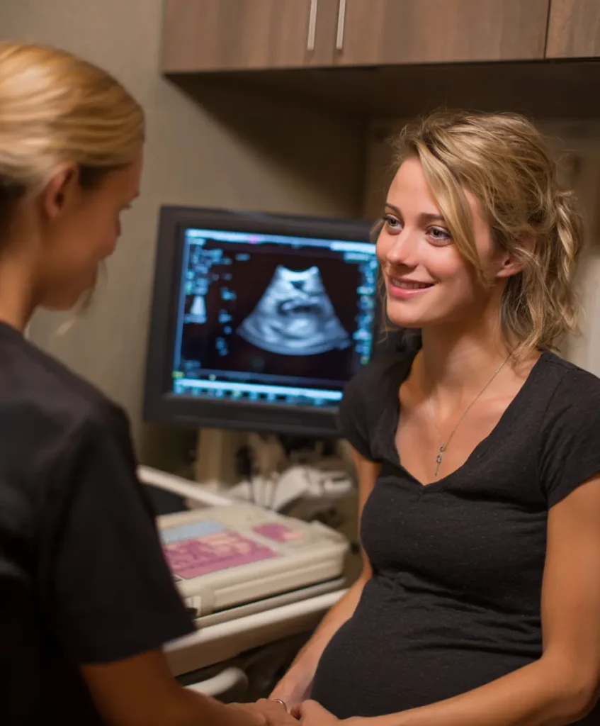
The 13 week pregnancy sonography is one of the most meaningful moments in early prenatal care. Occurring at the transition between the first and second trimester, this ultrasound scan provides crucial insights into fetal development, maternal health, and future pregnancy planning. At 13 weeks, the embryo has now developed into a fetus with recognizable human features, active movements, and significant organ formation.
This scan holds clinical and emotional importance: it reassures parents that the pregnancy is progressing normally, helps healthcare providers evaluate growth and anatomy, and establishes important baselines for future prenatal assessments. Whether this is a woman’s first pregnancy or part of her prenatal routine, the 13-week ultrasound offers clarity, connection, and valuable medical information.
Table of Contents
Why Is the 13 Week Pregnancy Sonography Done?
The 13 week pregnancy sonography serves both diagnostic and evaluative purposes. At this stage, the fetus has undergone rapid development, making it possible for clinicians to perform key measurements and screenings.
Confirming Gestational Age and Due Date
Although pregnancy dating is often established earlier, the 13-week scan allows for precise measurement of the crown–rump length (CRL), one of the most accurate indicators of gestational age. A corrected due date may be assigned if the measurement significantly deviates from previous estimates.
Assessing Fetal Development and Early Anatomy
At 13 weeks, the fetus is developed enough for clinicians to inspect major structures, including the skull, limbs, spine, abdomen, and thorax. Any early anomalies, although rare, may be identified.
Identifying Multiple Pregnancies
Twins or higher-order multiples can be confirmed at this visit, and their chorionicity and amnionicity—critical for managing twin pregnancies—can be determined with high accuracy.
Evaluating Placental Health and Amniotic Fluid
The placenta’s location, thickness, and general appearance are evaluated. Adequate amniotic fluid volume also provides reassurance that fetal kidneys are functioning and that pregnancy is progressing normally.
What Can Be Seen in a 13 Week Pregnancy Sonography ?
By 13 weeks, the fetus is about 7.4 cm long, roughly the size of a peach. Thanks to advancements in ultrasound technology, the 13 week pregnancy sonography allows for a wide range of detailed observations.
Visible Fetal Anatomy
- Head and facial structures: early profile, nasal bone, eye sockets
- Limbs and movement: arms and legs are fully formed; fingers and toes distinguishable
- Abdominal organs: stomach, bladder, and early bowel positioning
- Neural tube & spine: early assessment of spinal alignment
Fetal Movements
Even though the mother cannot feel them yet, the fetus is active. Movements may include:
- Stretching
- Yawning
- Thumb-sucking
- Bending and kicking
These help confirm healthy neuromuscular development.
Cardiac Activity and Blood Flow
The fetal heart beats between 140–170 bpm at this stage. Using Doppler, the sonographer may observe:
- Blood flow patterns
- Heart structure
- Early rhythm evaluation
Evaluation of the Maternal Cervix and Uterus
The cervix is observed to rule out shortening, funneling, or abnormalities. Uterine shape and any fibroids are documented for future follow-up.
Nuchal Translucency (NT) Screening during 13 Week Pregnancy Sonography
One of the most important parts of the 13 week pregnancy sonography is the Nuchal Translucency (NT) measurement. This test plays a central role in early screening for chromosomal abnormalities.
What Is NT?
NT refers to the layer of fluid-filled space behind the baby’s neck. At 13 weeks, increased fluid may indicate a higher risk for:
- Down syndrome (Trisomy 21)
- Edwards syndrome (Trisomy 18)
- Patau syndrome (Trisomy 13)
- Certain congenital heart defects
Ideal Timing
NT can only be accurately measured between:
- 12 weeks 5 days and
- 13 weeks 6 days
Beyond this window, the measurement is not valid.
Normal Range
A normal NT measurement is typically below 3 mm, although interpretation also depends on gestational age and fetal length.
Additional Factors Used in Risk Calculation
The NT measurement does not stand alone. A complete risk assessment includes:
- Maternal age
- Maternal weight
- Blood pressure
- Nasal bone presence or absence
- First-trimester blood test (PAPP-A, β-hCG)
Next Steps if Results Are Abnormal
An increased NT does not confirm a chromosomal anomaly; it simply indicates elevated risk. Further tests may be recommended:
- NIPT (Non-Invasive Prenatal Testing)
- CVS (Chorionic Villus Sampling)
- Amniocentesis
These provide more definitive answers about fetal chromosomal status.
Can Gender Be Detected at 13 Week Pregnancy Sonography ?
Although still early, determining fetal sex is sometimes possible during the 13 week pregnancy sonography.
Nub Theory
Between 11–14 weeks, the fetal genital tubercle (nub) may show a directional tilt:
- Upward angle (>30 degrees) → likely male
- Horizontal or downward angle → likely female
Accuracy can exceed 98% when the baby is in an optimal position, but incorrect angles or fetal movement may affect interpretation.
However, official confirmation is generally made during the 18–20 week anatomy scan, where visualization is clearer.

Preparing for the Ultrasound
Proper preparation improves image quality and ensures a smooth appointment.
Full Bladder
A partially full bladder helps lift the uterus out of the pelvis, providing better imaging—especially for women with a tilted uterus or early scans.
Hydration
Drinking water throughout the morning improves amniotic fluid clarity, enhancing fetal visibility.
Clothing
Loose, two-piece outfits make abdominal scanning easier.
Bring Essential Documents
- Prenatal record
- ID
- Requisition form
- Insurance card (if applicable)
Emotional Preparedness
Bringing a partner or support person may be reassuring. Prepare any questions you may want to ask the sonographer or doctor.
What to Expect During the Appointment
During the 13 week pregnancy sonography, the sonographer will follow standardized protocols to assess fetal and maternal structures.
Type of Scan
- Transabdominal scan is the standard.
- Transvaginal ultrasound may be used to improve NT accuracy or evaluate cervical length.
Procedure
- Gel is applied to the abdomen.
- A transducer is moved across the skin to capture images.
- Measurements are taken, including:
- CRL (Crown-Rump Length)
- NT thickness
- Fetal heart rate
- Images may be captured for medical records and parents.
Duration
Most scans last 45–60 minutes, depending on fetal position and the need for additional views.
Results
Some clinics provide results immediately, while others deliver reports within 1–3 days.

Emotional & Medical Significance of the 13 Week Pregnancy Sonography
The 13 week pregnancy sonography is more than a clinical examination—it is a milestone. For many parents, seeing the baby’s movements, hearing the heartbeat, and observing tiny details creates deep emotional bonding.
Medically, this scan provides the foundation for risk assessment, pregnancy planning, and ongoing prenatal monitoring. Early identification of abnormalities—though rare—allows for timely follow-up, genetic counseling, and appropriate decisions.
Conclusion
The 13 week pregnancy sonography is a key milestone that marks the shift into the second trimester and provides essential insight into both fetal and maternal health. This ultrasound helps confirm normal growth, evaluate early anatomy, assess chromosomal risk factors through NT screening, and establish the foundation for the rest of the pregnancy.
Beyond its medical importance, this scan offers reassurance and emotional connection, allowing parents to see their baby’s movements and development for the first time. With regular prenatal checkups, open communication with healthcare providers, and a good understanding of each pregnancy stage, women can make confident decisions that support a healthy pregnancy and a positive journey toward childbirth.
FAQs
1. How accurate is gender prediction on a 13-week ultrasound?
Gender prediction at 13 weeks can be up to 95–98% accurate when the fetus is in an ideal position and when the sonographer uses the nub theory. Accuracy depends on image clarity, fetal position, and technician experience.
However, the most reliable confirmation still occurs during the 18–20 week anatomy scan.
2. What is the difference between a girl and boy on a 13-week ultrasound?
At 13 weeks, the external genitalia are still developing, so sonographers use the angle of the genital nub:
Boy: The nub points upward (more than 30°) from the spine.
Girl: The nub is horizontal or slightly downward, parallel to the spine.
These differences are subtle, which is why gender prediction is not considered 100% certain at this stage.
3. What can the brain show in a 13-week ultrasound?
A 13-week ultrasound allows early visualization of major brain structures such as:
The developing forebrain, midbrain, and hindbrain
The choroid plexus
The early outline of the lateral ventricles
The posterior fossa
While detailed neuroanatomy is better assessed later, this scan checks for normal shape, symmetry, and e3. What can the brain show in a 13-week ultrasound?
A 13-week ultrasound allows early visualization of major brain structures such as:
The developing forebrain, midbrain, and hindbrain
The choroid plexus
The early outline of the lateral ventricles
The posterior fossa
While detailed neuroanatomy is better assessed later, this scan checks for normal shape, symmetry, and early structural development.
4. At 13 weeks pregnant, can you tell the gender by ultrasound?
Yes, gender can often be suggested at 13 weeks using the nub theory, especially during high-resolution scans. However:
It is not guaranteed
Accuracy depends on fetal position and ultrasound quality
Official confirmation is usually done at 18–20 weeks
So gender can sometimes be predicted at 13 weeks, but it should be considered early and not definitive.
5. What do the legs look like on a 13-week ultrasound?
On a 13-week scan, fetal legs are clearly visible and usually show:
Fully formed long bones (femur, tibia, fibula)
Active movement such as kicking or stretching
Feet with distinguishable toes
Symmetry between both legs
These observations help confirm healthy limb development and overall fetal growth.
Written by a Gynecologist based on medical experience and scientific evidence to help you make informed decisions about 13 Week Pregnancy Sonography